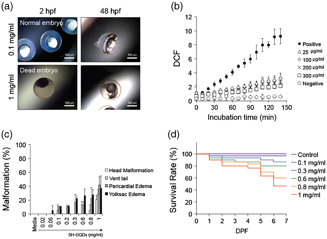
Fig. 6.

(a) Photographs of zebrafish embryos exposed to SH-GQDs (0.1 and 1 mg/ml). (b) Quantitative measurements of DCF fluorescence using multimode detector (Ex: 485 nm; Em: 515 nm). Embryos were treated with H2O2 (50 μM) in presence of varying concentration of SH-GQDs (0, 25, 100, 200 and 300 μg/ml) (n = 3). (c) Malformation of zebrafish embryos exposed to SH-GQDs (n = 20). (d) Survival analysis of varying concentrations of SH-GQDs (n = 30).
