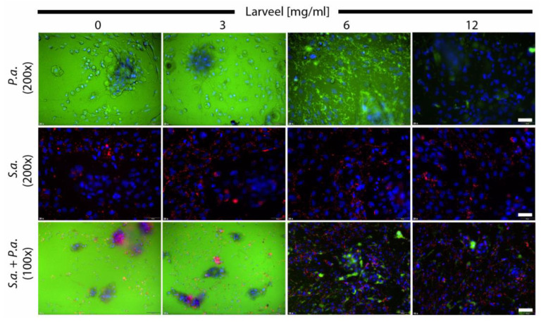
Figure 1.
Fluorescence microscopy of cells infected with bacteria. Fibroblast and keratinocyte co-cultures were infected with P. aeruginosa and S. aureus in medium with 0.5% antibiotics and were supplemented with different Larveel® concentrations for 24 h. P. aeruginosa is shown in green fluorescence (gfp), S. aureus in red fluorescence (rfp) and nuclei of dermal cells are shown in blue (DAPI). Nuclei of keratinocytes were arranged in clusters. P.a.: P. aeruginosa; S.a.: S. aureus. Top and middle line 200× magnification, scale bar: 50 µm; bottom line 100× magnification, scale bar: 100 µm.

