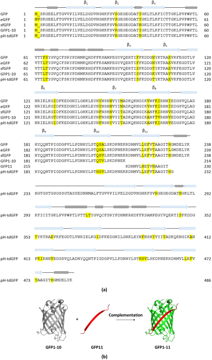
Figure 2.
(a) Sequence alignment and structural annotation of GFP, eGFP, sfGFP, splitGFP and pH-tdGFP. Helices are indicated in grey and β-strands as blue arrows. Differing amino acid identities in the alignments are highlighted in yellow. (b) Split-GFP complementation: separating the 10 N-terminal β-strands (GFP1–10) from the 11th β-strand (GFP11) results in the generation of split-GFP that becomes fluorescent upon complementation (Created in PyMOL).

