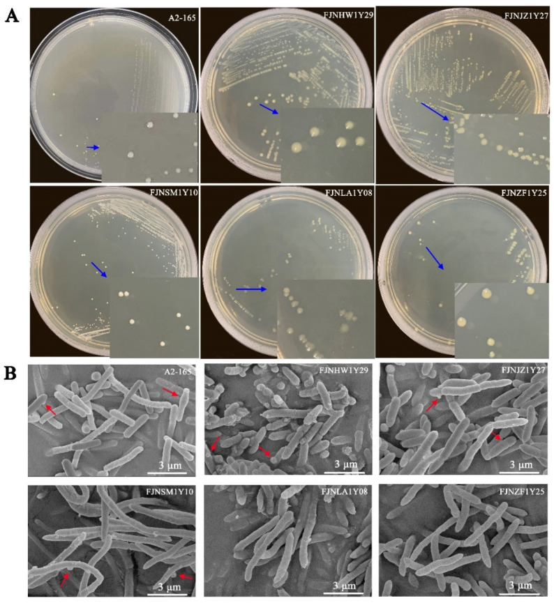
Figure 2.
Colony and scanning electron microscopy images of F. prausnitzii strains: (A) the colony images of F. prausnitzii isolates and (B) the scanning electron microscopy images of F. prausnitzii isolates. The blue arrows show that colonies of F. prausnitzii isolates were 2–4 mm in diameter, circular, and opaque to transparent. The red arrows indicate the special ultrastructure of “swelling”. Scale bars indicate 3 μm.

