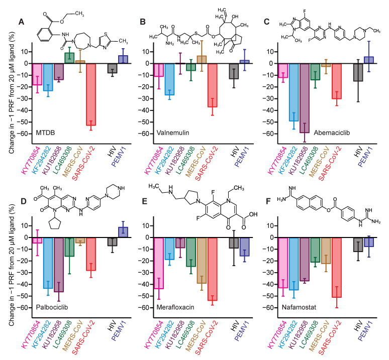
Figure 4.
Activity of −1 PRF inhibitors against frameshift signals from different CoVs. (A) Change in −1 PRF efficiency compared to basal levels (Figure 2) induced by 20 μM MTDB. Remaining panels show the same for (B) valnemulin, (C) abemaciclib, (D) palbociclib, (E) merafloxacin, and (F) nafamostat. In each case, results for CoVs are shown on left, results for specificity controls on right. Experiments performed in vitro using dual-luciferase reporter in rabbit reticulocyte lysate. Error bars represent standard error of the mean from 3–6 replicates. Insets: chemical structures of inhibitors.

