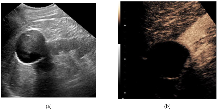
Figure 1.
A 79-year-old man with a history of bladder cancer undergoing evaluation for hydronephrosis. Grayscale ultrasound image of the left kidney in the longitudinal orientation (a) shows an exophytic hypoechoic mass containing internal low-level echos. Following an intravenous injection of 1.8 cc Lumason ultrasound contrast, a contrast-enhanced ultrasound image focused at the upper pole (b) revealed the mass was completely non-enhancing (devoid of signal), which is diagnostic for a simple cyst. No further follow-up was necessary.

