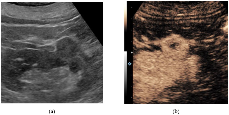
Figure 2.
A 67-year-old man with multiple renal lesions, status post SBRT one year prior for contra-lateral RCC. Grayscale ultrasound image of the left kidney in longitudinal orientation (a) shows an isoechoic exophytic nodule. Following the intravenous administration of 1.0 cc Lumason ultrasound contrast, a contrast-enhanced ultrasound image (b) shows the nodule demonstrating predominantly solid avid enhancement relative to the adjacent renal cortex. A partial nephrectomy revealed clear cell renal cell carcinoma, grade 2.

