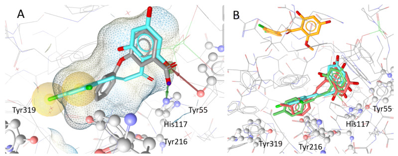
Figure 6.
Localisation and orientation of chalcones to AKR1C3 in docking simulations: (A) docking pose of the protein (PDB Code: 3UWE) with its co-crystallised ligand 3-phenoxybenzoic acid (gray; IC50 = 0.68 µM) and compound 23 (cyan), the compound with the highest inhibitory effect. The hydrophobic interactions between compound 23 and Tyr319 and Tyr216 are highlighted in yellow. The red and green arrows indicate hydrogen bonds between the hydroxy group of ring B of 23 and Tyr55 and His117, respectively; (B) shows the docking poses of the highly inhibiting chalcones 23 (cyan), 20 (red), and 21 (green) in comparison with weak inhibitor 25 (orange), which was placed outside the binding pocket in simulations.

