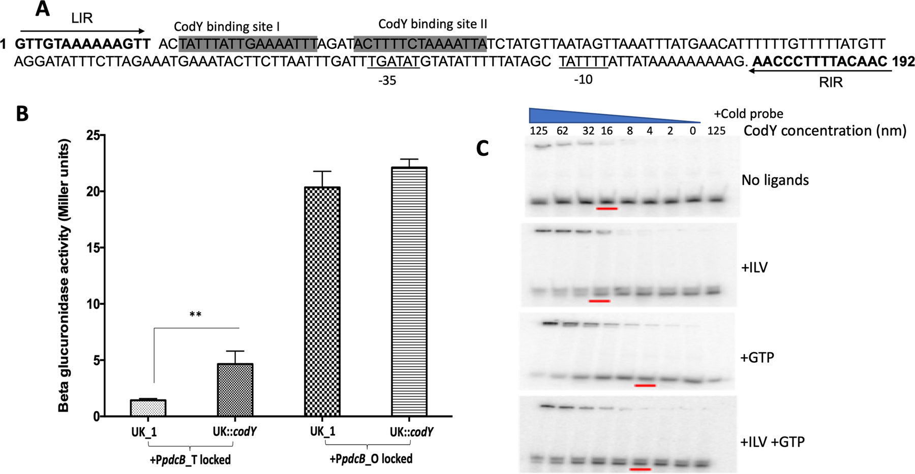
Figure 6. CodY represses the expression of pdcB, which is partially relieved by DNA inversion.

A. Schematic showing the potential CodY binding sites that lie within the invertible region upstream of pdcB. LIR and RIR are in bold and marked with arrows B. Beta-glucuronidase activity of PpdcB-gusA fusions locked in either translucent or opaque orientation in the parent UK1 and UK1::codY mutant. The data shown are the standard errors of the mean of three biological replicates. Statistical analysis was performed using two-way ANOVA.** indicates p<0.05.C. Binding of purified CodY to the upstream region of pdcB. Binding was increased with increasing concentration of CodY. The presence of GTP and ILV further enhanced CodY binding. Ten-fold excess of the cold probe was added with 125 ng of purified CodY in a control reaction to rule out non-specific binding.
