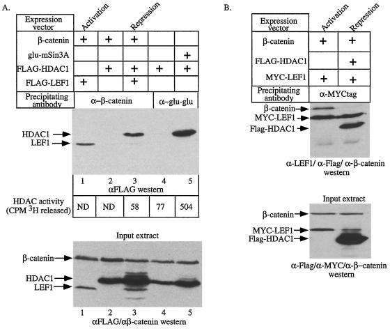
FIG. 4.
β-Catenin can interact with HDAC1 and attenuate HDAC activity. (A) 293 cells were transfected with expression vectors encoding the indicated cDNAs. The amount of FLAG-tagged LEF1 or FLAG-tagged HDAC1 associated with β-catenin or mSin3A in each extract was detected by Western blotting for the FLAG epitope following immunoprecipitation with antisera specific for β-catenin or the Glu-Glu epitope on mSin3A (top panel). The values at the bottom of the top panel are the HDAC activities determined for each immunoprecipitation. ND, not determined. Western blotting of whole-cell extracts shows that the transfected HDAC1, LEF1, and β-catenin proteins are expressed to similar levels (bottom panel). The top and bottom panels are aligned so that the IPs shown in a given lane were performed from the cell extracts shown in the lane directly below. (B) 293 cells were transfected with expression vectors encoding the indicated cDNAs. The proteins associated with MYC-LEF1 were detected with a mixture of antibodies specific for the FLAG epitope, LEF1, and β-catenin (top panel). Western blotting of whole-cell extracts shows that the transfected HDAC1, LEF1, and β-catenin proteins are expressed to similar levels (bottom panel). The panels are aligned as in panel A. In both panels, “Activation” and “Repression” mark the lanes showing experiments performed under activation and repression conditions, respectively.

