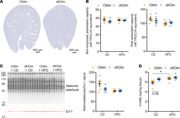
Figure 7. Analysis of the antioxidant capacity and level of lipid peroxidation in cKOm mice fed under HFD.
(A) Gross morphological structure and Oil Red O staining of neutral lipid depositions (in red) in the kidney of cKOm and Ctrlm adult mice fed under HFD for 4 weeks. The absence of the red color indicates the absence of lipid deposition. (B) Results of a Trolox assay performed on renal extracts showing nonenzymatic antioxidant capacity (left panel) and total antioxidant capacity (right panel) of Ctrlm and cKOm mice fed under control diet (CD) or HFD for 4 weeks. (C) Immunoblot performed on renal extracts targeting the product of lipid peroxidation malondialdehyde (left panel) and its quantification (right panel) in Ctrlm and cKOm mice fed under CD or HFD for 4 weeks. DJ-1 immunoblot is used as a loading control to normalize malondialdehyde abundance. (D) Amount of 4-HNE measured by competitive ELISA in kidney extracts from Ctrlm and cKOm mice fed under CD or HFD for 4 weeks. Box and whiskers represent mean ± SEM. Two-way ANOVA and post hoc Tukey’s multiple comparisons test, *P < 0.05.

