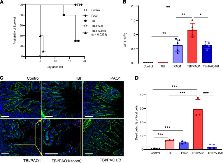
Figure 1. PAO1 alters the survival of irradiated mice.
(A) C57BL/6J mice after TBI (9.25 Gy) were infected with PAO1 in presence or absence of lipoxygenase inhibitor (baicalein) and monitored for more than 15 days. Unirradiated (C) and unirradiated PAO1-infected (PAO1) mice served as controls. Data are pooled from 2 independent experiments. (B) PAO1 burden in colon. Fecal samples were collected, resuspended, and serially plated on cetrimide agar and incubated at 37°C for 20 hours. PAO1 colonies were counted and represented as CFU/g. Data represent mean ± SD, n = 5 mice/group. **P < 0.01; *P < 0.05, 1-way ANOVA, Tukey’s multiple comparison test. (C) Baicalein treatment mitigated epithelial barrier disruption induced by TBI plus PAO1. Damage was assessed as discontinuity of the actin layer (green), and cell death (pink nuclei) that was particularly evident at the apex of the crypt. Dead cell (red), F-actin (green), and Hoechst (blue). Scale: 50 μm. (D) Baicalein treatment mitigated cell death induced by TBI plus PAO1. TBI, total body irradiation; PAO1, P. aeruginosa; B, baicalein. Data represent mean ± SD, n = 5 mice/group for control, TBI/PAO1/B; n = 3, for TBI, PAO1, and TBI/PAO1. ***P < 0.001, 1-way ANOVA, Tukey’s multiple comparison test.

