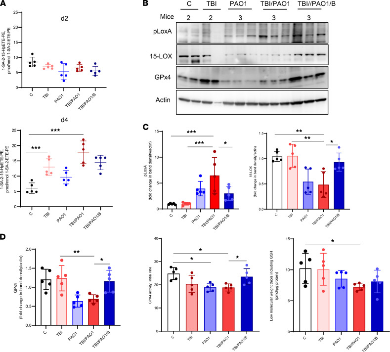
Figure 4. PAO1 enhances radiation-induced ferroptosis.
(A) Levels of proferroptotic signal 1-stearoyl-2-15(S)-HpETE-sn-glycero-3-phosphoethanolamine (1-SA-2-15-HpETE-PE) were higher in irradiated mice after PAO1 infection. 24 hours after TBI, mice were left alone (TBI) or infected with PAO1 in the absence (TBI/PAO1) or presence of baicalein (TBI/PAO1/B). Ileum samples were collected on days 2 and 4 after TBI and processed for redox-lipidomics. Data represent mean ± SD, n = 5 mice/group; ***P < 0.0001, 1-way ANOVA, Tukey’s multiple comparison test. (B) Presence of 15-LOX, pLoxA, and GPx4 in ileum. Ileum homogenate from day 4 after radiation was tested for presence of 15-LOX, pLoxA, and GPx4 using antibody against 15-LOX and pLoxA. (C) Densitometry-based quantitative assessment of 15-LOX and pLoxA protein. Mean relative intensity of pLoxA (left) or 15-LOX (right) was normalized to actin. Data are mean ± SD, n = 5 mice/group; ***P < 0.0001; **P < 0.001; *P < 0.01, 1-way ANOVA, Tukey’s multiple comparison test. (D) Assessment of GPx4 protein and activity from day 4. Densitometry-based quantitative assessment of GPx4 (left). Mean relative intensity of GPx4 was normalized to actin. Data are mean ± SD, n = 5 mice/group; **P < 0.001; *P < 0.01, 1-way ANOVA, Tukey’s multiple comparison test. GPx4 activity measurements (middle). Ileal homogenates (15 μg of protein) after TBI were incubated in buffer containing 0.1 M Tris (pH = 8.0), 0.5 mM EDTA, 1.25% Triton X-100 with 1SA-2-15-HpETE-PE (30 μM), glutathione (150 μM), glutathione reductase (1 U/mL), and NADPH (35 μM). Activity was monitored by the disappearance of NADPH fluorescence (excitation wavelength: 340 nm; emission wavelength: 460 nm). Data are mean ± SD, n = 5 mice/group; *P < 0.05, 1-way ANOVA, Tukey’s multiple comparison test. Low-MW thiols and GSH content in gut homogenates on day 4. For calculation of GSH, fluorescence of Thiol probe IV disappearing after treatment with GPX were used. Data are mean ± SD, n = 5 mice/group; *P < 0.01 versus control, 1-way ANOVA, Tukey’s multiple comparison test.

