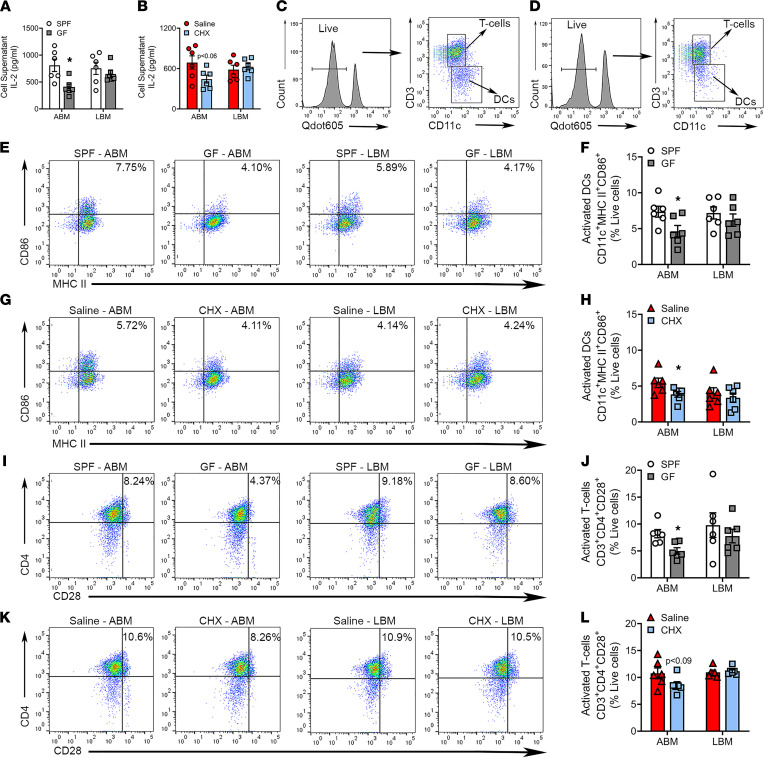
Figure 8. Commensal oral microbiota promotes activation of alveolar BM–derived DCs in vitro.
(A and B) DC/T cell allostimulation assay supernatants were collected for ELISA IL-2 analysis from (A) SPF versus GF alveolar BM (ABM) and long BM (LBM) cultures and (B) saline versus CHX ABM and LBM cultures. (C and D) Representative gating strategy for T cells and dendritic cells from (C) ABM and (D) LBM cultures. (E and F) Representative dot plots of final gating and quantitative measures for CD11c+MHC II+CD86+ activated DCs from ABM and LBM cultures derived from SPF versus GF mice; reported as percentage of live cells. (G and H) Representative dot plots of final gating and quantitative measures for CD11c+MHC II+CD86+ activated DCs in ABM and LBM cultures derived from saline versus CHX mice; reported as percentage of live cells. (I and J) Representative dot plots of final gating and quantitative measures for CD3+CD4+CD28+ activated T cells in ABM and LBM cultures derived from SPF versus GF mice; reported as percentage of live cells. (K and L) Representative dot plots of final gating and quantitative measures for CD3+CD4+CD28+ activated T cells in ABM and LBM cultures derived from saline versus CHX mice; reported as percentage of live cells. Unpaired t test; data presented as mean ± SEM; *P < 0.05.

