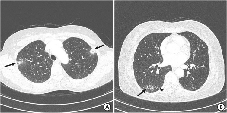
Figure 1. Chest CT scans obtained from patients with trastuzumab deruxtecan-induced lung injury. (A) A CT image obtained from a 60-year-old woman with organizing pneumonia in the upper lobes (case 1 in Table 2) shows peripheral, multifocal, nodular consolidations (arrows). The patient developed drug-induced interstitial lung disease 331 days after beginning treatment with trastuzumab deruxtecan; however, the lung abnormalities remained even after glucocorticoid therapy. (B) A CT image obtained from a 53-year-old woman with organizing pneumonia in the right lower lobe (case 4 in Table 2) who received her first dose of trastuzumab deruxtecan 407 days previously showed focal consolidation and ground-glass opacity (arrow) adjacent to underlying fibrosis (arrowhead). The patient’s condition improved after cessation of trastuzumab deruxtecan without glucocorticoid therapy.
CT = computed tomography.

