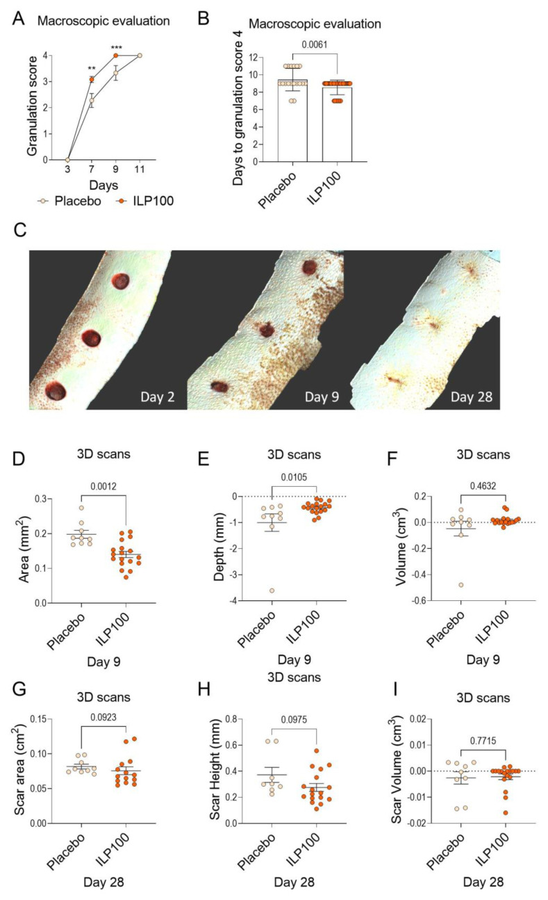
Figure 4.
Wound healing assessed by macroscopic evaluation and from 3D scans in Cohort B. Wounds in Cohort B were macroscopically evaluated for granulation tissue formation by granulation score day 3 to 11 (A) and days to granulation score 4. ** p < 0.01, *** p < 0.001 (B) (Placebo: n = 3, N = 18, ILP100: n = 6, N = 36). The healing was also assessed using 3D scans, where panel (C) shows representative projections of 3D scans from day 3, day 9, and day 28. From the 3D scans, wound area, wound depth, and wound volume were measured (D–F) for placebo and ILP100-treated wounds day 9 (Placebo: n = 3, N = 9, ILP100: n = 6, N = 18). On day 28 when the wounds were healed, scar area, scar height, and scar volume (G–I) were measured from 3D scans, (Placebo: n = 3, N = 9, ILP100: n = 5–6, N = 14–18). Statistic comparison between the groups were made using Mann-Whitney tests (A,B,D–I).

