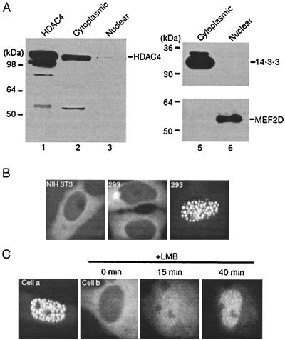
FIG. 1.
Cytoplasmic localization of HDAC4. (A) Affinity-purified Flag-HDAC4 (lane 1) and cytoplasmic (lanes 2 and 5) and nuclear (lanes 3 and 6) extracts of NIH 3T3 cells were subjected to immunoblotting with the anti-HDAC4 (lanes 1 to 3), anti-14-3-3 (lanes 5 and 6, top), or anti-MEF2D (lanes 5 and 6, bottom) antibody. The amount of extracts was normalized according to cell numbers. The 55-kDa band in lane 2 may not be specific, since it was not reproducibly detected by different bleeds of the anti-HDAC4 antibody. (B) Representative green fluorescence images of NIH 3T3 and 293 cells expressing GFP-HDAC4. (C) Green fluorescence images of two SKN cells (cells a and b) expressing GFP-HDAC4. After initial examination for green fluorescence, LMB (10 ng/ml) was added to the medium and cell b was then analyzed for redistribution of green fluorescence at the indicated times. Under similar conditions, LMB had minimal effects on the pancellular localization of GFP itself (data not shown).

