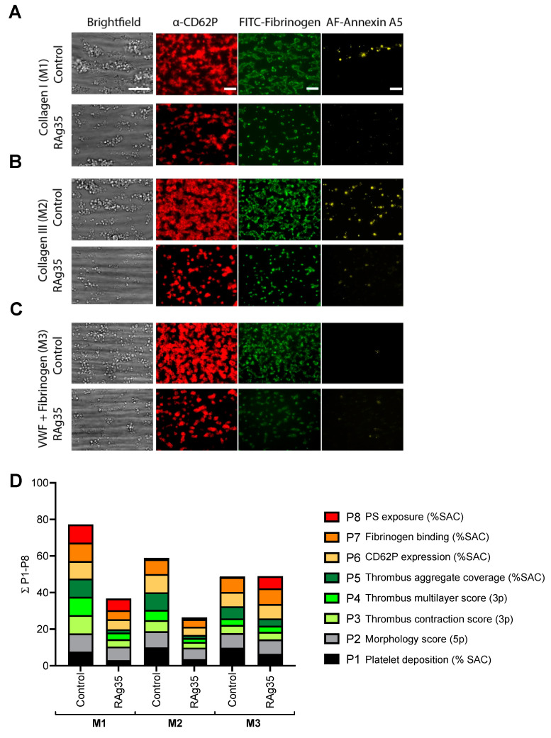
Figure 3.
Thrombus and aggregation-reducing effect by blockage of the interaction of GPIbα with VWF A1 domain at high-shear flow. Whole-blood was preincubated for 10 min with CLB-RAg35 mAb (RAg35, 10 µg/mL) or equal volume of saline. Blood samples were flowed over microspots consisting of collagen-I (M1), collagen-III (M2), or VWF/fibrinogen (M3) for 3.5 min at wall-shear rate of 1600 s−1. (A–C) Representative brightfield microscopic images at end stage. Bars = 20 µm. Brightfield images were used for analysis of adhesion-related parameters, namely platelet deposition (P1) and morphological score (P2), and aggregation-related parameters (P3–5). End-stage three-color fluorescence images used for platelet activation assessment: CD62P expression (AF647 anti-CD62P mAb, P6), fibrinogen-binding (FITC anti-fibrinogen mAb, P7), and phosphatidylserine exposure (AF568 annexin A5, P8). (D) Cumulative representation of scaled values (0–10) per parameter. Color reflects adhesion parameters P1–2 (shades of black), aggregation parameters P3–5 (shades of green), and activation parameters P6–8 (shades of red). For details, see Figure S3.

