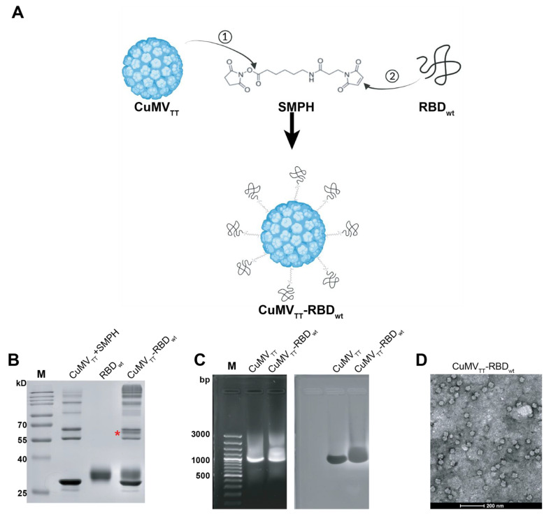
Figure 1.
Generation and characterization of CuMVTT-RBDwt. (A) Scheme of covalent reactions between CuMVTT, RBDwt and SMPH. (B) SDS-PAGE gel analysis of CuMVTT-RBDwt. The coupled CuMVTT-RBDwt band (56.3kD) was marked with a star. (C) Examination of RNA contents inside CuMVTT-RBDwt comparing with CuMVTT VLP (left); the same agarose gel was stained with Coomassie, illustrating the protein bands located at the same position as RNA bands (right). (D) Transmission electron microscope image of CuMVTT-RBDwt, which demonstrates sphere viral shape.

