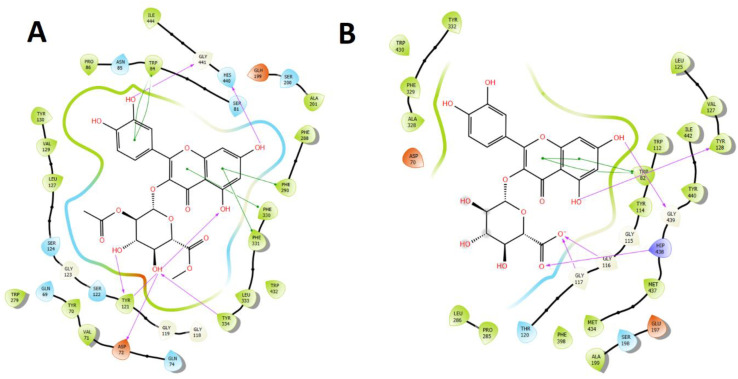
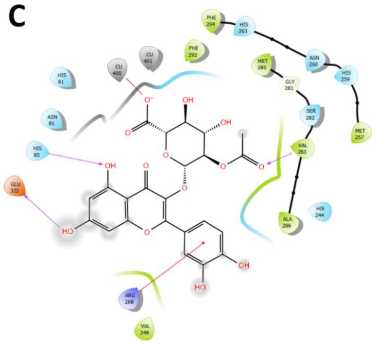
Figure 8.
Two-dimensional diagram of (A). Most active compound: Quercetin 3-O-ß-D-2″-acetylglucuronide methyl ester (related to docking binding energy) and its main interactions into the acetylcholinesterase (TcAChE) catalytic site. (B). Most active compound: Quercetin 3-O-(ß-D-glucuronide) (related to docking binding energy) and its main interactions into the butyrylcholinesterase (hBuChE) catalytic site. (C). Most active compound: Quercetin 3-O-ß D-2″-acetylglucuronide (related to docking binding energy) and its main interactions into the tyrosinase catalytic site. Purple arrows represents hydrogen bond interactions, green lines represents π-π interactions and red lines represents π-cation interactions.


