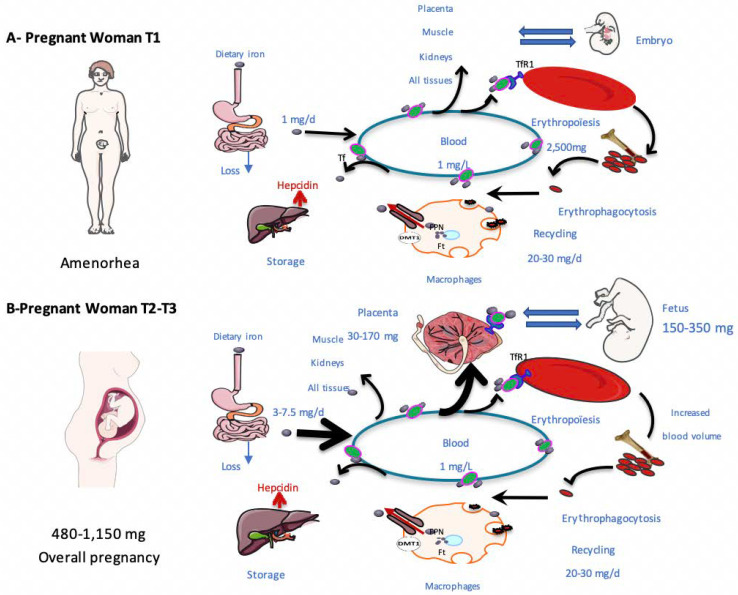
Figure 1.
Schematic description of iron metabolism during pregnancy. This figure describes systemic iron metabolism during the first (A) and the second (B) part of pregnancy. Iron is presented as grey circles and transferrin as green circles. The average quantities of iron in the various processes are mentioned. Dietary iron is absorbed by enterocytes. Iron is distributed to various tissues and cells via transferrin in the plasma. It is internalized in tissues by endocytosis via transferrin receptors (TFR1). In pregnant women, placental TFR1 can import transferrin from the maternal circulation into the placenta through the syncytiotrophoblast. Bone marrow captures 70% of plasma iron for hemoglobin synthesis in hematopoietic precursors. At the end of their life, erythrocytes are phagocytosed by macrophages. The liver plays a significant role in iron storage. Hepcidin is synthesized by the liver and can contribute to the internalization and degradation of FPN at the basal side of enterocytes and on the macrophage membrane. During the second part of the pregnancy, the iron need increased. Tf: transferrin, TFR1: transferrin receptor 1, FPN: ferroportin.

