Figure 3.
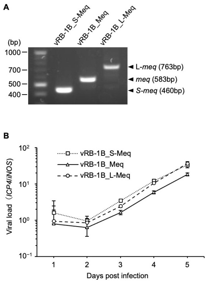
Expression analysis and replication of recombinant MDVs in cell culture. (A) mRNA expression of each meq-isoform in chicken embryo fibroblasts (CEFs) infected with vRB-1B_S-Meq, vRB-1B_Meq, and vRB-1B_L-Meq was confirmed by reverse transcription-polymerase chain reaction (PCR). (B) CEFs were infected with 50 plaque-forming units of recombinant Marek’s disease viruses. The infected cells were collected daily for 6 days. The viral loads in the infected cells were analyzed by quantitative PCR. The growth kinetics among the groups were analyzed using the Kruskal–Wallis test.

