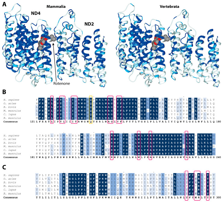
Figure 2.
(A) Residue conservation mapped on the structure of rotenone bound ND4 and ND2 subunits (PDB ID 6ZKM) for Mammalia (left panels) and Vertebrata (right panels). The ribbon color goes from white (0% conservation) to light blue (70%), medium blue (90%), and dark blue (100%). The rotenone is reported in a space-fill representation, colored according to the atom type. (B,C) Multiple alignment and conservation analyses of ND4 and ND2 protein regions binding rotenone. Alignments of protein sequences from mammals, including Homo sapiens used as the reference sequence, are reported. Different shading corresponds to increasing conservation levels: amino acid conservation between 70% and 90% are highlighted in light blue, amino acid conservation between 90% and 99% are highlighted in medium blue, and invariant positions (100% conservation) are highlighted in dark blue. Alignment gaps are indicated by a dash (-). Amino acids involved in rotenone binding are boxed in pink, while those undergoing conformational changes in LHON mutant are boxed in yellow.

