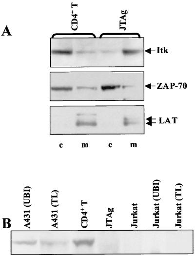
FIG. 1.
Constitutive localization of Itk to the membrane is correlated with absence of PTEN expression. (A) The cytosolic (c) and membrane (m) fractions prepared from normal human CD4+ T cells and JTAg T cells were immunoblotted with a monoclonal antibody to Itk (2F12). The membrane was stripped and reblotted with rabbit antisera to the cytosolic protein ZAP-70 (1213) and the transmembrane protein LAT (3023). (B) Whole-cell lysates (total protein, approximately 20 μg for JTAg and Jurkat E6 cells, and 10 μg for CD4+ T cells, A431 cells, and Jurkat [Upstate Biotechnology Inc. {UBI}] and Jurkat [Transduction Laboratories {TL}] cells) were immunoblotted with an antibody cocktail of four anti-PTEN antibodies (see Materials and Methods). The two A431 lysates (from UBI and TL) were included as positive controls.

