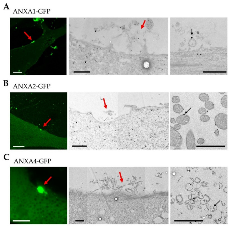
Figure 4.
CLEM imaging of ANX in damaged LHCN myotubes. ANXA1-GFP (A), ANXA2-GFP (B), or ANXA4-GFP (C) expressing LHCN myotubes were damaged by laser ablation (red arrow) and immunostained for ANX using a secondary antibody coupled to gold nanoparticles (black particles). Fluorescence images obtained about 90 s after laser ablation are presented (left-hand image) together with TEM images (middle and right-hand images). Respective middle and right-hand images have been collected from different sections. Right-hand images show ANX (black particles) interacting with circular lipid structures (black arrow). Scale bar for fluorescence images: 10 µm; for TEM: 1 µm.

