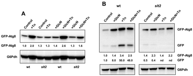
Figure 7.
HBP activation and SLT2 knock-out causes distinct responses to autophagy. (A) Overnight SCD-Leu grown cultures of pRS415-GFP-ATG8 transformants of the BY4741 wild-type (wt) and slt2 strains were refreshed in YPD (OD600 = 0.1) lacking or containing 11.5 mM glucosamine (GlcN) and grown at 30 °C until OD600 ~ 0.3. Aliquots were withdrawn for their immediate analysis (Control), and cultures were split in two and incubated at 30 °C in the presence (Tn; GlcN+Tn) or absence (control; GlcN) of 2 μg/mL tunicamycin (Tn) for 3 h. Protein extracts were prepared as described in Section 2, separated by SDS-PAGE, and analyzed by Western blot for GFP-Atg8 and free GFP using anti-GFP antibody. The image shows only a part of the gel where GFP-Atg8 was localized as free GFP was hardly detected. (B) The same strains were assayed as above except that the tunicamycin (Tn) treatment was extended for 6 h. In all cases, the bands corresponding with GFP-Atg8 or free GFP (GFP) are indicated. The level of glucose-6-phosphate dehydrogenase (G6Pdh) was used as a loading control for crude extracts. The values at the bottom of the images represent the GFP-Atg8 and free GFP abundance relative to that of the wild-type strain under control conditions that was set at 1.0. Representative experiments are shown.

