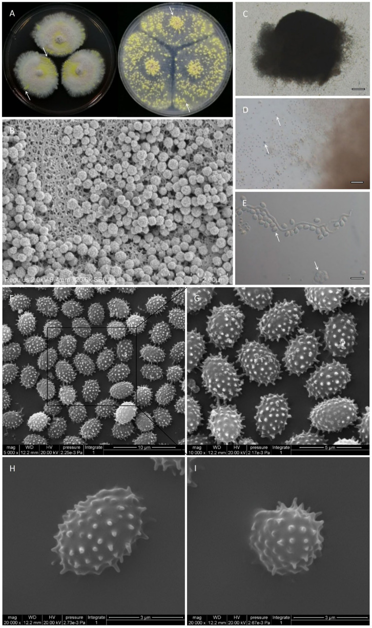
Figure 7.
Teleomorphic stage of JP-NJ4 (CCTCC M 2012167). (A) Colonies inoculated for 2 wk on CZ (left) and OA (right). (B–I) Micromorphological characters of JP-NJ4. (B) Primary ascomata collected from OA (inoculation for 1 wk), observed by scanning electron microscope (SEM). (C–D) A mature ascoma that is releasing asci and ascospores at different magnification ((C) 5×; (D) 20×), observed by optical microscope (Zeiss). (E) An ascus and ascospores (100×). (F–I) Ascospores, observed by SEM. Scale bars: B = 2 μm; C = 200 μm; D = 50 μm; E = 10 μm; F = 10 μm; G = 5 μm; H = 3 μm; I = 3 μm.

