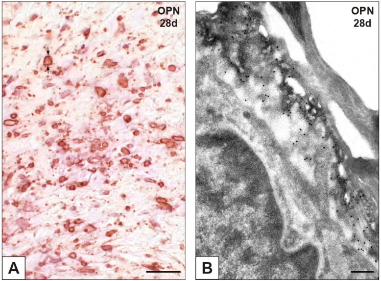
Figure 5.
Immunohistochemical detection of osteopontin (OPN) in aortic valve leaflets implanted into rat subcutis for 28 days (28d). (A) Histological section showing positive valvular cells exhibiting marked reactivity at their edges (counterposed arrows). (B) Thin section subjected to immunogold labelling showing a membrane-derived pericellular layer decorated by gold particles. Bar: 0.25 mm (A); 0.25 μm (B).

