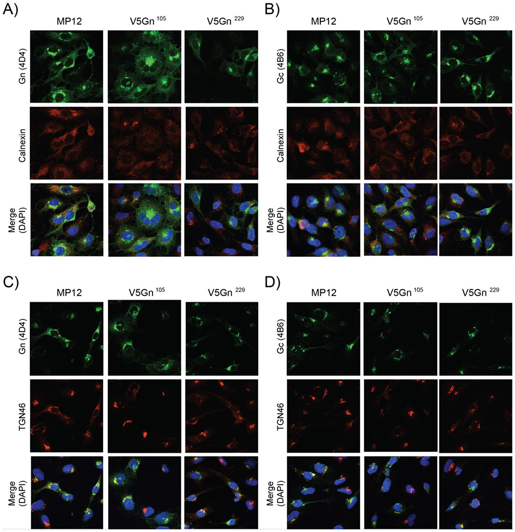
Figure 2. Cellular localization of Gn and Gc proteins.

HSAECs cells were infected with either the parental MP12 or V5-tagged viruses at a MOI 1. At 24 hpi, cells were then fixed and analyzed by immunofluorescence for Gn (panels A, C) or Gc (B, D) protein localization. Calnexin (A, B), TGN46 (C, D), and DAPI staining were utilized as controls for the endoplasmic reticulum (ER), Golgi, and the nucleus, respectively.
