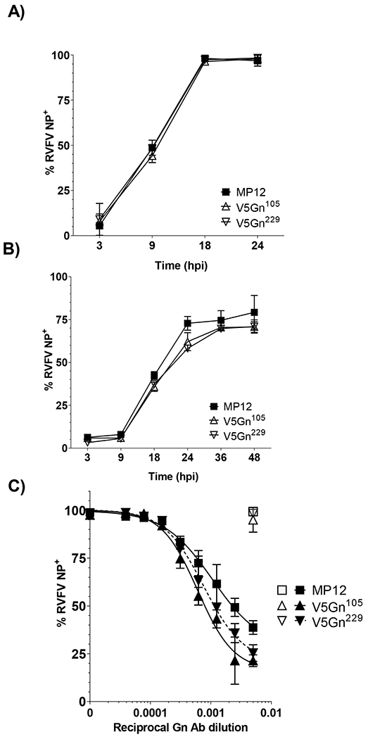
Figure 3. Impact of Gn V5 insertion on viral spread and neutralization.

A) Huh-7 and B) mosquito C6/36 cells were infected at a MOI 0.1 with parental MP12 or V5-tagged viruses. Viral spread over time was assessed by RVFV NP staining and flow cytometry. Both mean and standard deviations are plotted for three biological replicates. C) RVFV virus inoculum (MOI 0.1) was mixed with increasing 2-fold dilutions of Gn antibody and incubated on Vero cells for 24h. After 48 hpi, cells were harvested and the percentage of RVFV positive cells were determined by flow cytometry. Filled symbols represent data from virus inoculum mixed with Gn antibody. Open symbols represent data from virus inoculum mixed with NP antibody at 1:200 dilution. Data is graphed as a percentage relative to no antibody control. Both mean and standard deviations are plotted for three biological replicates and non-linear regression analysis was performed. The EC50 for MP12 is a 935-fold dilution, while the EC50 for V5Gn105 and V5Gn229 is a 1649-fold and a 1314-fold dilution, respectively.
