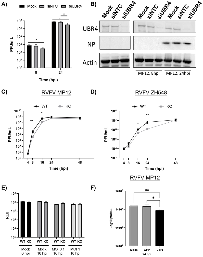
Figure 5. Absence of UBR4 decreases RVFV viral titers.

A-B) UBR4 expression in Huh-7 cells was knocked down by siRNA treatment (50 nM). After 72h, cells were infected with MP12 (MOI 0.1). At 8 and 24 hpi, infectious viral titers (A) or protein expression of UBR4, Actin, and RVFV Gn and NP were examined (B). C-D) HEK 293T WT and UBR4 KO cells were infected with untagged parental RVFV attenuated strain, MP12, (C) or the fully virulent strain, ZH548, (D) at a MOI of 0.1. Viral supernatants were collected at various time points post infection and titers were determined by plaque assay. E) HEK 293T WT and UBR4 KO cells were either mock-infected or infected with the parental MP12 virus (MOI 0.1 or 1). Cellular viability was measured either prior to infection or at 16 hpi using Cell Titer-Glo. F) Loss of Ubr4 decreases RVFV replication in mosquito cells. CxQx cells were plated in a 24 well plate and transfected with 100ng of dsRNA per well. The transfected cells were infected with RVFV MP12 (MOI 0.01). The supernatants were collected and viral titer determined by plaque assay. Mock-transfected control cells, GFP- dsRNA transfected cells; and Ubr4- dsRNA transfected cells. Figures 5 A and C–F represent the mean and standard deviation of three biological replicates. *p-value<0.05, **p-value<0.01.
