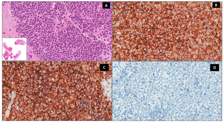
Figure 2.
Histologic findings. A predominately dermal blue nodule composed of small–medium-sized round cells with a round nucleus, fine granular chromatin, inconspicuous nucleoli, and scanty cytoplasm (A) ×200, Lower insert ×20 hematoxylin-eosin, (H&E). Immunohistochemically, the neoplastic cells are positive for CD56 (B) ×200 and synaptophysin (C) ×200 and negative for chromogranin A (D) ×200.

