
Figure 5.
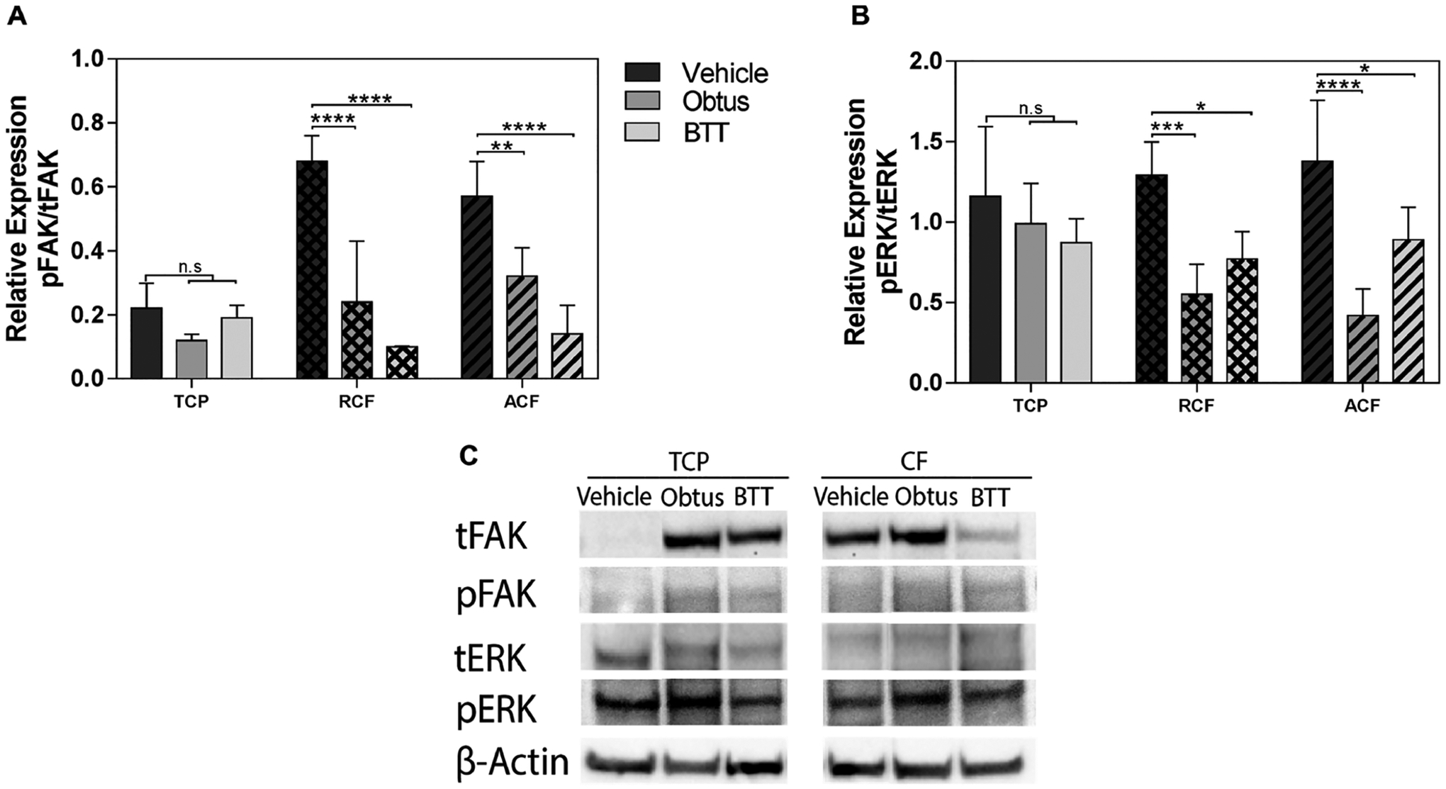
FAK and ERK signaling activity. Quantification of relative expression of total focal adhesion kinase (tFAK; 119 kDa) and phosphorylated FAK (pFAK; 119 kDa), total extracellular-signal-regulated kinase (tERK; 42–44 kDa), and phosphorylated ERK (pERK; 42–44 kDa) in tumor cells relative to β-actin (42 kDa). Quantification of (A) pFAK in tFAK expression and (B) pERK in tERK by image analysis of the Western blots bands. Data represent the relative mean intensity ± standard error of three independent experiments. (C) Representative protein bands are shown for TCP and collagen fibrils (CF).