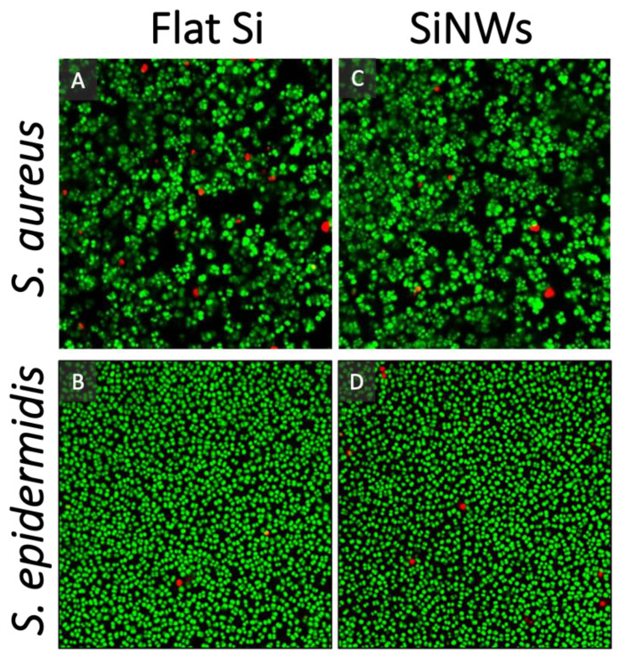
Figure 4.
Representative Live/Dead Confocal Scanning Laser Microscopy (CSLM) images for Staphylococcus spp. stained with Syto9 (green, live) and propidium iodide (red, dead) cultured for 24 h over: (A,B) Flat silicon; and (C,D) SiNW surfaces. Field of view is 71.8 µm × 71.8 µm. The confocal images were taken at the interface between the surfaces and the biofilm, showing only the first layer of cells in contact with the surface.

