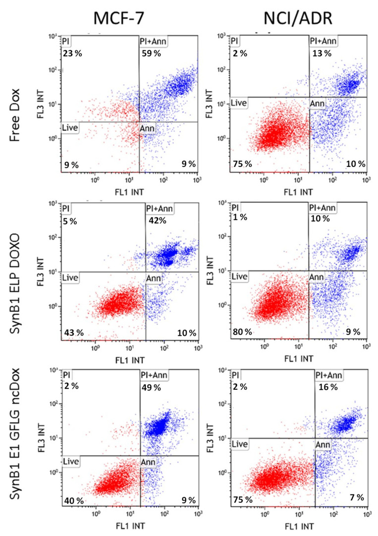
Figure 7.
Induction of apoptosis by free dox, SynB1-ELP-DOXO, and SynB1-ELP-GFLG-ncDox in MCF-7 and NCI/ADR cell lines. Scatter plots show the live (lower left quadrant), early apoptosis (lower right quadrant), late apoptosis (upper right quadrant), and necrotic (upper left quadrant) MCF-7 and NCI/ADR cells after they were treated for 24 h with 2 µM dox-equivalent drug concentration. Percentage of apoptotic cells was determined based on gating for double staining with PI and Annexin V Alexa488.

