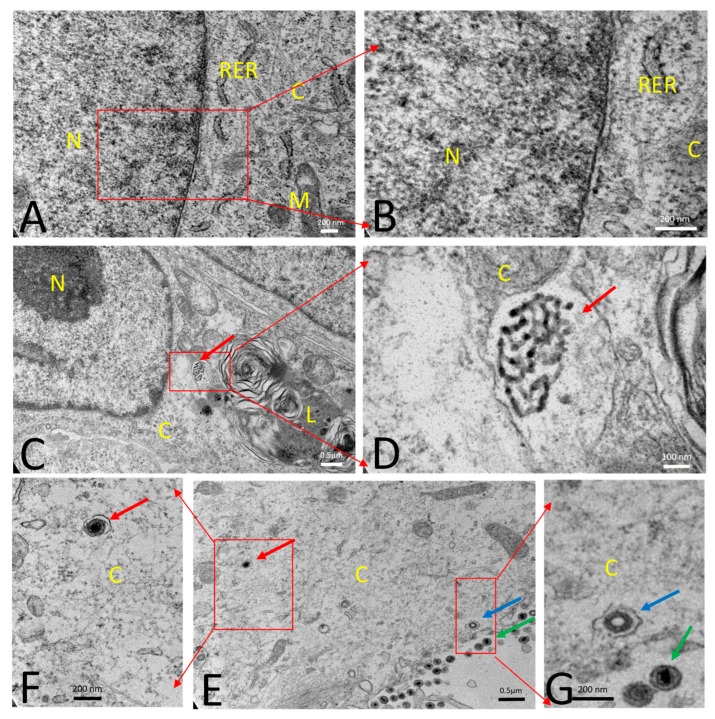
Figure 5.
Transmission electron microscopy: (A,B) Mock infected healthy swine kidney (SK) cell with normal cellular morphology. (C,D) SK cells were infected with porcine circovirus type 2b (PCV2b) (infected at an MOI of 0.1) and fixed at 72 h post-infection (hpi). PCV2b infected cell showed accumulation of PCV2 viruses (red arrow) within the vesicle-like structures in the cytoplasm. Each PCV2 particle measured about 20 nm in diameter. (E–G) SK cells were infected with PRV wt (infected with an MOI of 5) and fixed at 12 hpi. PRV wt-infected cells showed enveloped herpesvirus (red arrow; about 200 nm in diameter) within the vesicle-like structure in the cytoplasm. The process of budding and release of several enveloped viruses were also noticed on the periphery of the cell near the plasma membrane (blue arrow). Released virus particles from the outer surface of the cells were accumulated in intercellular space (green arrow). (B, D, F, G) are magnifications of (A, C, E), respectively. N—Nucleus; C—Cytoplasm; RER—Rough endoplasmic reticulum; M—Mitochondria; L—Lysosomes.

