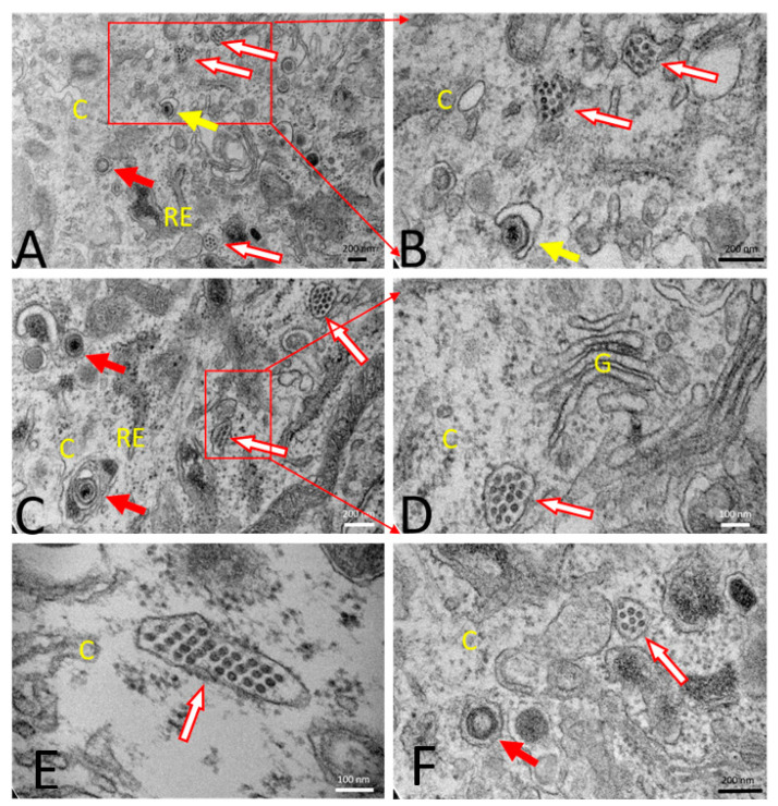
Figure 6.
Transmission electron microscopy. (A) PRVtmv+ vaccine virus-infected swine kidney (SK) cells (infected at an MOI of 5) were fixed at 18 h post-infection. PRVtmv+ infected cells showed several fully enveloped 200 nm in diameter size PRVtmv+ vaccine virus particles in the exocytic vesicles in the cytoplasm (red arrows). Secondary envelopment of intracytoplasmic PRVtmv+ capsids by budding into vesicles was visible (yellow arrows). Most of the PRVtmv+ infected SK cells showed several accumulations of PCV2 virus-like particles (VLPs) within the vesicular structures in the cytoplasm (red arrow with a white fill). The PCV2-VLPs were circular, and each measured about 20 nm in diameter. (B) Magnification of A is given. (C) The presence of enveloped PRVtmv+ vaccine virus (red arrow) and PCV2 VLPs within vesicles (red arrow with a white fill) in the cytoplasm of the PRVtmv+ infected SK cells were visible. (D–F) Accumulations of PCV2-VLPs within the vesicle-like structures in the cytoplasm of the infected cells (red arrow with a white fill). C—Cytoplasm; RER—Rough endoplasmic reticulum; G—Golgi apparatus.

