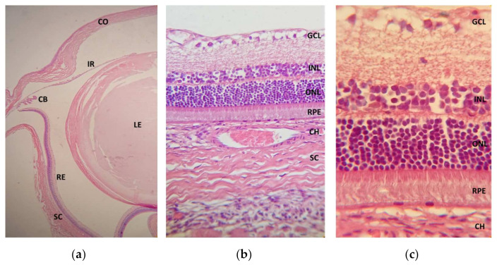
Figure 10.
Photomicrographs of the treated eye cross-sections after CS/HA-EPOβ administration stained with hematoxylin and eosin: (a) ocular globe from group C (magnification 4×); CO, cornea; IR, iris; CB, ciliary body; LE, lens; RE, retina; SC, sclera. (b) cross-section from a rat’s retina from group D (magnification 40×); CH, choroid; GCL, ganglion cell layer; INL, inner nuclear layer; ONL, outer nuclear layer; RPE, retinal pigment epithelium. (c) cross-section from a rat’s retina from group E (magnification 100×).

