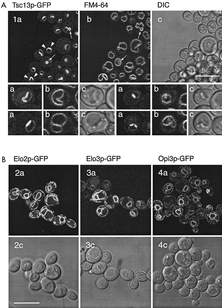
FIG. 9.
(A) C-terminally tagged Tsc13p-GFP shows a typical ER localization pattern and, in addition, is highly enriched in a region of the nuclear envelope adjacent to the vacuole. Selected cells are shown at higher magnification in the lower panel. (B) Elo2p-GFP, Elo3p-GFP, and Opi3p-GFP localize to the ER; i.e., around the nucleus and at the cell periphery. a, GFP fluorescence; b, FM4-64-labeled vacuolar membranes; c, DIC transmission images. Each scale bar is 10 μm.

