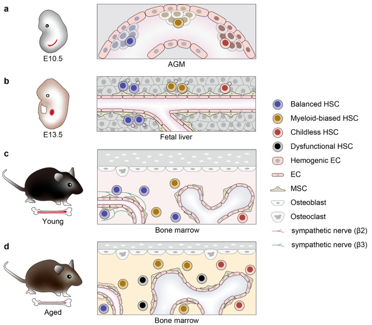
Figure 2.
Clonal expansion of functionally distinct HSCs during development and aging. (a) Development of HSCs from hemogenic endothelium of the AGM region in mouse embryos at E10.5. HSCs arising from different regions of the AGM can be considered as distinct clones and are shown in different colors. (b) At around E13.5, HSCs migrate to fetal liver and clonally expand by self-renewing cell division, while keeping their lineage potential. The degree of expansion differs for each clone [7], and expansion of HSCs with balanced lymphoid and myeloid potential could possibly be favored by the fetal liver niche [52]. (c) After birth, HSCs further migrate to bone marrow and most of them enter quiescence in periarteriolar (left) or perisinusoidal (right) HSC niche. (d) During aging, HSCs are exposed to various cell-intrinsic and -extrinsic stressors that selectively expand myeloid-biased HSC clones and also induce functionally defective HSC clones, leading to myeloid-biased hematopoiesis and impaired capacity of hematopoietic regeneration. EC, endothelial cells; MSC, mesenchymal stromal cells.

