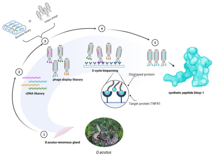
Figure 5.
Schematic description of the established workflow for the screening of DAvp-1. (1) Extraction of venom glands from D. acutus. (2) mRNA was extracted from the venom glands of D. acutus and transcribed into a cDNA library. (3) Construction of a phage display library using the cDNA library of D. acutus. (4) Coating TNFR1 on the ELISA plate and performing three rounds of screening towards a D. acutus. venom phage library. (5) The screened phages were sequenced, and a series of analyses were performed to obtain DAvp-1.

