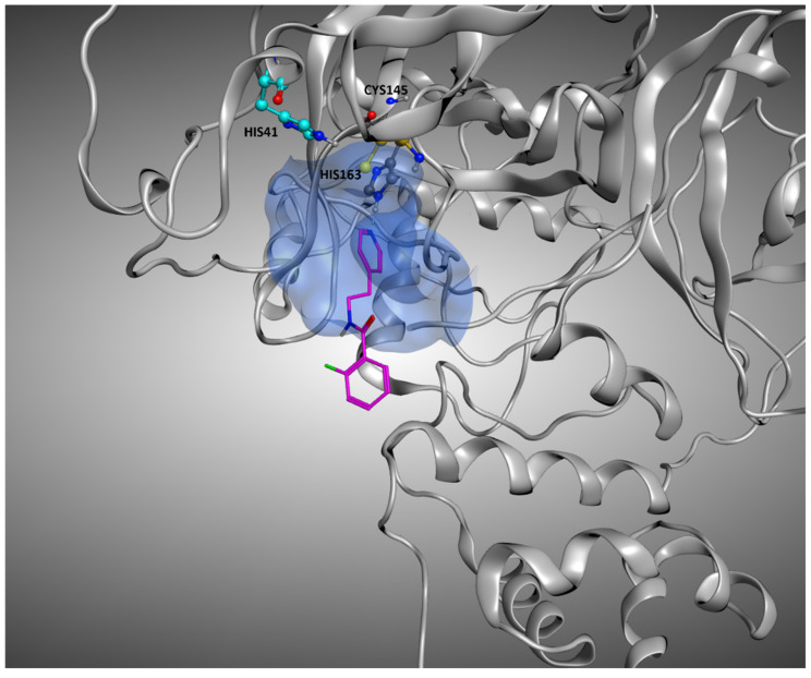
Figure 11.
Representation of the crystallographic complex conformation of 5RGK, one of the protein–ligand complexes in which the crystallographic ligand is located inside the orthosteric binding site, but the docking calculation results in high RMSD values. This is mainly due to the high level of solvent exposure that characterizes this ligand, which locates just a small portion of its structure inside the pocket, leaving the rest in an outer zone. The ligand is represented with stick representation (C-atom are colored in magenta), and the catalytic dyad (Cys145 and His41) is highlighted, as well as the His163 and the binding site residue interacting with the ligand. To give a better representation, the surface of the protein in a 5 Å radius from the ligand is represented and colored in blue.

