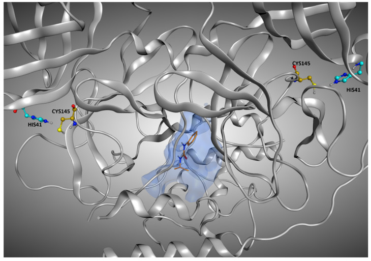
Figure 12.
Representation of the crystallographic pose of 7LFP, which is one of the protein–ligand complexes in which, even if the crystallographic ligand is located outside the orthosteric binding site, the RMSD values between the original coordinates and the ones given from the docking runs are considerably low. The reason for this can be found in the very low solvent exposure of this ligand, which is located in the interface between the monomers, and so is shielded by them. The ligand is represented in orange, and the catalytic dyad (Cys145 and His41) of both monomers is highlighted. To give a better representation, the surface of the protein in a 5 Å radius from the ligand is represented and colored in blue.

