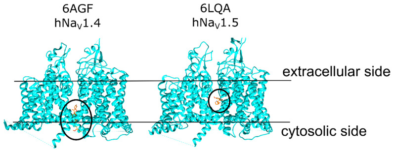
Figure 2.
The structures of human NaV1.4 (PDB ID: 6AGF) bound to a detergent molecule at the “mouth site” (left), and the human NaV1.5 (PDB ID: 6LQA) bound to quinidine at the “pore site” (right). The ligand molecules are shown in orange for both structures. The approximate region of each binding site is marked with a black circle. The black bar represents the putative position of the membrane with the extracellular and cytosolic sides of the membrane labelled.

