Figure 1.
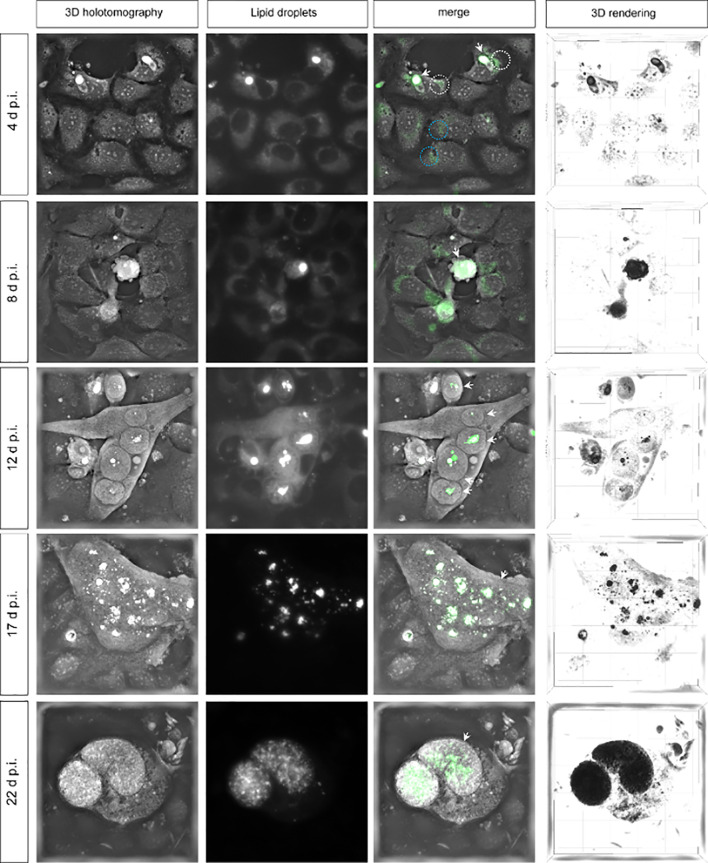
Lipid droplet formation in Eimeria bovis-infected host cells. Live-cell 3D analyses of E. bovis-infected host cells revealed the presence of an increasing number of lipid droplets during macromeront formation. 3D holotomography represents refractive index (RI) images; lipid droplets are stained by LipidSpot™ (green); merges show co-localization of white structures with higher RI and LipidSpot™-stained lipid droplets (green). 3D rendering shows lipid droplets in black in a 3D model. White arrows indicate E. bovis stages; ×600 images.

