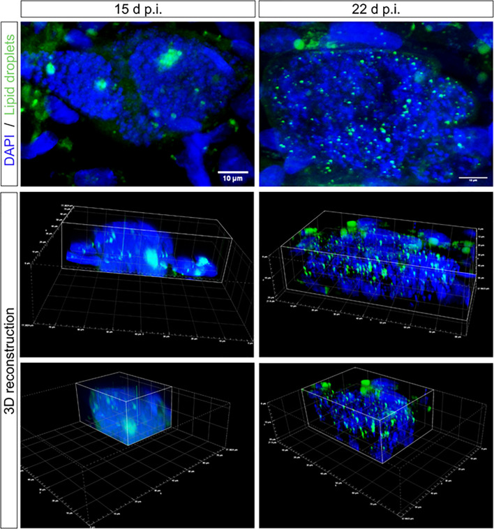
Figure 2.
Lipid droplet distribution in E. bovis macromeronts. Confocal microscopy analysis evidenced the presence of large lipid droplets being positioned as large clusters in immature macromeronts at day 15 p.i. In contrast, in mature macromeronts (22 d p.i.), numerous small lipid droplets (green) were evenly distributed in the parasitic compartment as also illustrated by 3D reconstructions. DAPI (blue) was used to stain DNA. Scale bar 10 µm.

