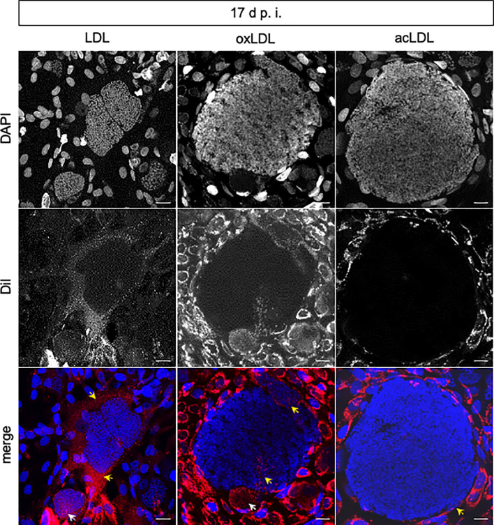
Figure 3.
Internalization of LDL, acLDL, and oxLDL by E. bovis-infected host endothelial cells and meronts I. Confocal analyses revealed that Dil-LDL, Dil-oxLDL, and Dil-acLDL (red) were internalized into the host cell cytoplasm at all time points studied (n = 3, representative illustration). Intrameront LDL and oxLDL internalization (white arrows) was observed at 17 d p.i. In contrast, acLDL was not observed within meronts I. DAPI (blue) stained DNA. Internalization of marked lipids in the cytoplasm of host cells (LDL, oxLDL, acLDL) or stretches of the parasitophorous vacuole membrane (oxLDL) are signed with yellow arrows. Scale bar 20 µm.

