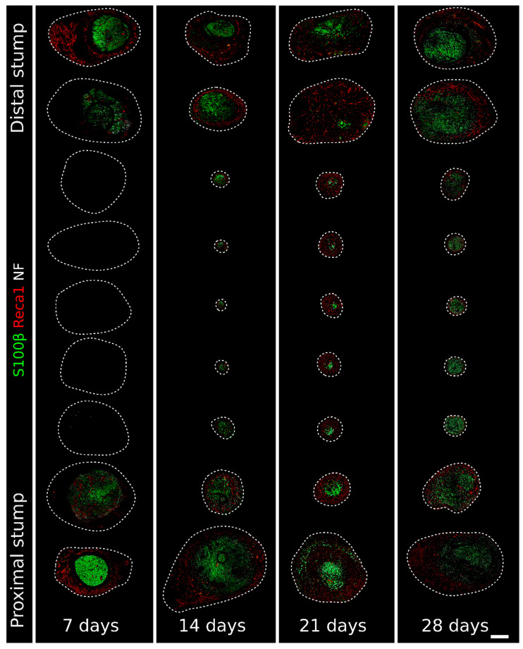
Figure 2.
Immunofluorescence staining of regenerating nerves 7, 14, 21, and 28 days after the injury and repair. One section every millimeter labeled with Reca1 (red, endothelial cell marker), S100β (green, Schwann cell marker), and Neurofilament/NF (white, axon marker) to follow the nerve regeneration progression. The single labeling is shown in Figures S1 (NF), S2 (S100β), and S3 (Reca1). The dotted line delimits the region containing cell nuclei identified with DAPI (as in Figure 1B). Scale bar: 400 µm. It is possible to zoom in on the high-resolution version of this figure to appreciate the interactions between the different structures. Available online: https://zenodo.org/record/6198513 (accessed on 30 November 2021).

