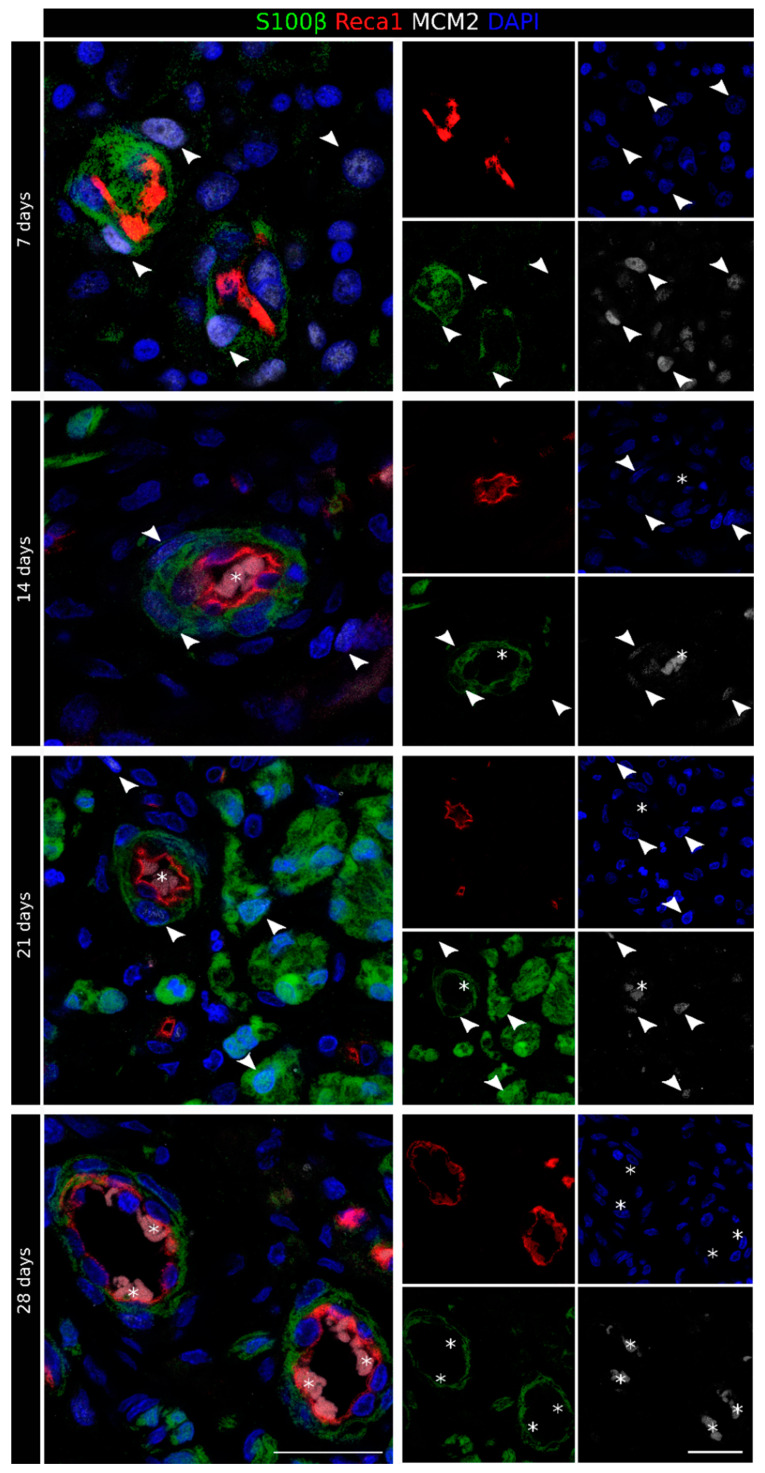
Figure 4.
Identification of newly formed Schwann cells in the regenerating nerve. Proliferation marker staining of the regenerating nerve 7, 14, 21, and 28 days after the injury and repair. For each time point, the merged picture is shown on the left, while the four single labeling samples are shown on the right. Sections were labeled with MCM2 (white, proliferation marker), S100β (green, Schwann cell marker), Reca1 (red, endothelial cell marker), and DAPI (blue, nuclear marker). Erythrocyte autofluorescence was detected inside some blood vessels (identified by an asterisk). MCM2-positive Schwann cells are indicated using an arrowhead. Scale bar: 25 µm.

