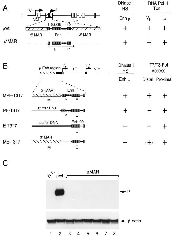
FIG. 1.
Schematic diagram of transgenes containing μ enhancer sequences. (A) Structure of the rearranged μ wild-type and ΔMAR transgenes containing the intragenic enhancer between the rearranged VDJ and Cμ exons. The positions of the VH and Iμ promoters are shown by arrows. The intragenic μ enhancer (Enh), including the Iμ promoter and binding sites for transcription factors (gray boxes) (20), is flanked by MARs (hatched boxes). (B) Structure of T3T7 transgenes in which μ enhancer sequences are linked to bacteriophage T3 and T7 promoters (arrows) at enhancer-proximal and -distal (1 kb) positions (33). The MPE-T3T7 gene construct contains the entire μ enhancer fragment (Enh), which includes the Iμ promoter (P) and enhancer core (E), and a single flanking MAR (M). The PE-T3T7 gene contains the Enh fragment without MAR sequences. The construct E-T3T7 contains only Enh 90, and the construct ME-T3T7 contains the Enh 90 fragment and a single MAR. DNA fragments (1 kb) from the large T (LT) and VP1 genes of simian virus 40 acted as reporters of transcription from the T3 and T7 promoters (33). “Stuffer” sequences replace μ LCR fragments in order to maintain the spatial relationship of the transgene components. To the right of each transgene (A and B), the data obtained by Forrester et al. (23) and Jenuwein et al. (33) are summarized. The formation of DNase I-hypersensitive sites (HS) at the μ enhancer and the generation of transcripts (VH and Iμ) by endogenous RNA polymerase II (Pol II Txn) or transcripts (T7, distal; T3, proximal) by exogenous bacteriophage RNA polymerases (T7/T3 Pol Access) are indicated by a plus sign. (C) Total RNA samples isolated from the B-cell cultures used in the in vivo footprinting experiments were probed for the presence of VH-initiated transcripts by S1 nuclease protection assay. Detection of the β-actin mRNA was used as an internal control. As expected from previous data (23), the transgenic enhancer in all μ ΔMAR cell lines did not activate the distal VH promoter at a detectable level (lanes 3 through 8). The total RNA from nontransgenic pre-B cells (N.T.; lane 1) and from μ-transgenic pre-B cells (μwt; lane 2) were used as controls.

